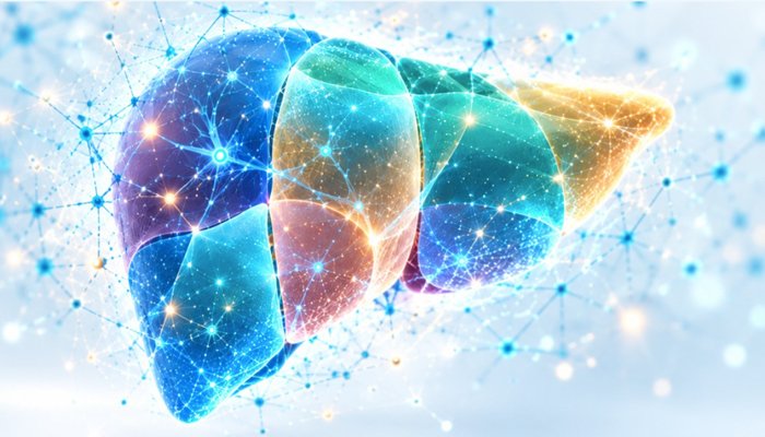
Building in the Open: The Start of LiverRa
Production Diary, Entry 1: Turning a clinical research script into a deployable diagnostic tool.
By Levan Gogichaishvili, MD
TAKEAWAY: LiverRa aims to provide an objective, standardized marker of early disease progression, earlier than standard two-dimensional assessment. Using AI tools to support clinical decisions, not replace them. We are seeking partners for development.
I. The Limits of Two-Dimensional Measurement
Routine liver imaging still relies largely on two-dimensional measurements to assess a three-dimensional organ.
The standard clinical workflow plays out the same way in hospitals around the world. A patient with a history of liver disease gets an abdominal contrast CT. The liver is measured on an axial slice, a 12-centimeter width is recorded, and mild surface irregularity is noted. By standard criteria, the report is correct, and the organ appears stable.
But a damaged liver doesn’t deteriorate uniformly along a neat geometric axis. Often before laboratory values or standard measurements become clearly abnormal, the organ remodels asymmetrically. As fibrosis progresses, the caudate lobe, which has its own venous drainage, often enlarges while the right lobe shrinks. The spleen enlarges as portal pressure rises.
When these changes become obvious on standard measurements, the disease may already have reached advanced cirrhosis, or a malignant tumor may have developed. Catching that morphologic shift early requires measuring changing volumetric ratios across the entire organ in 3D, an analysis that is impractical to perform by hand across hundreds of imaging slices.
II. The Heavy Lifting: Cohorts and Volumetrics
The urgency of this project comes from the specific clinical frontline we are operating on.
In our region of the Caucasus we are managing a unique cohort of patients. Thanks to a successful national public health program, tens of thousands of local patients have had their hepatitis infections completely cured. The viral load is gone. But the structural tissue damage left behind remains a risk. They are biologically cured, but architecturally compromised.
To address the wide range of compromised patients and increased diagnostic complexity and workload we have dedicated ourselves to an building an original system with three core components:
- The Neural Net: A computer vision pipeline that automates CT segmentation across hepatic tissue, vasculature, and pathology to generate an anatomically precise organ digital twin.
- The Measurement Module: An automated analytics layer coupled directly to the segmentation model that tracks contrast agent dynamics to profile lesion enhancement patterns and calculate cross-organ volumetric ratios.
* Note: Tissue pathology is a timed perfusion sequence, not a static snapshot. The module must benchmark density across baseline, arterial wash-in, and 7-to-10-minute delayed equilibrium phases. Tracking the clearance rate exposes the vascular signatures that distinguish benign tissue remodeling from early malignancy and is missing in many diagnostic training models today.
- The Report Writer: A clinical language model that synthesizes multi-phase perfusion data and volumetric ratios into standard radiological reporting syntax, generating an initial structured diagnostic draft in minutes.
The Report Writer closes the mechanical circuit, tying the digital twin and contrast metrics directly into standard clinical syntax. By chaining all three modules together, the pipeline eliminates the tedious drudgery of manual slice-tracing and mental arithmetic. What used to burn hours on a PACS workstation is reduced to an actionable draft in minutes, clearing the queue without cutting procedural corners.
Automated generation, however, is never an autonomous diagnosis. The machine constructs the draft, but the physician holds the pen and signs the chart. This is strictly a computational stethoscope—an instrument designed to surface quantitative ground truth, cut down cognitive fatigue, and get out of the way so the clinician can make the final call.
III. Bringing Clinical Experience into Tool Design
LiverRa grew out of our combined surgical practice within the Liver Research Network at GeoHospitals in Tbilisi, where liver damage found in the operating room was often more extensive than standard two-dimensional measurements suggested. The clinical reality under the retractor did not always match the measurements on the screen. The project has two goals: to reduce diagnostic errors where advanced care is available, and to improve the quality and safety of care where expert specialists and modern software tools are scarce.
Diagnostic software has traditionally been developed by dedicated engineering teams working with licensed hospital data. This model has produced many valuable tools. Its limitation is distance from the bedside: the people writing the algorithms are rarely the people who operate on the organ, and details that matter in surgery can be missed.
Wider access to AI computing and open-source medical software has changed who can take part. Clinicians can now contribute directly to building diagnostic tools. They can be developed by a practicing surgeon closing the clinic door at five o’clock, pulling open-source medical software onto a workstation, and wrestling with Python code until midnight.
When surgeons and engineers work together, the result is easier to use in practice. The tool reflects real anatomy, not an idealized CAD model, because the parameters are set by someone who knows exactly where the resection margin lies.
IV. Why build a Network to build a product?
For all the benefits of clinical experience and workflow, aspiring independent, AI-supported diagnostic tool developers have two main problems:
First: Clinical experience makes a tool more useful in practice, but a single expert working alone soon reaches a limit.
A lone practitioner can write the script, curate a local dataset, and build a working prototype. What one clinician cannot provide alone is multi-center validation, international peer review, and the long-term support needed for clinical adoption. You cannot stress-test a diagnostic model across fifty different health systems from a single desk. If you build a tool in a vacuum, it will carry the biases of your local scanners and your specific patient demographics.
Second: The greatest hurdle to model success is ADOPTION. A developer must get other experts to use, review and promote the new device or procedure. Isolated independent entrepreneurs have little or no ability to access the level of attention required to spur widespread use.
Dr. Levan Gogichaishvili
Diagnostic AI is maturing, but adulthood looks less like an AI revolution and more like a thousand niche tools that quietly make clinicians faster or more consistent—and only the ones with proof and payment stick.
Prognosis:Innovation
These are the exact structural gaps the Liver Research Network was built to close.
LiverRa is being developed within the network rather than as a standalone project for a number of reasons. The network brings together surgeons, radiologists, hepatologists, engineers and data scientists, to assist with advice, peer review and data.
It serves as a real-time validation framework for ongoing work product to provide valuable developer feedback and guidance while also fostering an active network of reliable clinical early adopters.
More broadly, it provides the collaborative infrastructure to turn an isolated script into a resilient, field-tested reality, pairing the builder’s clinical ground truth with an active, shared ecosystem that provides the scale no single doctor can generate alone.
V. Scaling the Blueprint and Seeking Partners
A working script on a local hard drive is a great proof of concept, but it isn’t a deployable clinical tool. The next mechanical step is pure scale.
We were looking for a way to grow this project from one surgeon’s workstation into a team effort, so on behalf of the Liver Research Network we applied to NVIDIA Inception, the NVIDIA program for startups. In late September we were accepted.
Membership brings a range of benefits: discounts on NVIDIA software, training through the NVIDIA Deep Learning Institute, go-to-market support, and the opportunity to apply for cloud computing credits and GPU access through partners such as Google Cloud, among many others.
Most importantly for us, it provides a working environment and tools that allow many people to take part in a single project remotely, from anywhere in the world, and opens large reserves of support for those who want to help build the product we are aiming for. Inception also connects member startups with NVIDIA’s network of venture capital firms through its VC Alliance, opening a path to investors who understand AI.
One hundred cases are now fully processed. Models for the arterial and delayed phases are already trained, building on pretrained open-source models but now our own, and their results match manual outlining almost perfectly. Training of the venous phase, the most difficult one, is under way. This phase is meant to support the planning of most treatment modalities, including surgery and other interventions. The first target is a model trained on 1,000 cases drawn from a much larger archive. That expansion is what will allow us to lock in the statistical thresholds for our volumetric ratios.
Our network allows interested researchers and clinicians from different countries, working in very different circumstances, to put their local resources and expertise to use in the scientific and clinical research carried out within the network. Every project provides valuable entrepreneurial experience and access to income opportunities with global institutions. For those who commission research, this is also a major opportunity: within a single network, they will gain access to a global pool of talent and potential partners.
Finally, a model trained at a single center may not meet the workflow or operational considerations for other institutions and patient populations. So active feedback, beta testing and work product validation will be a large proportion of our engagement opportunities as well.
To this end, both to garner feedback for our project and to act as a blueprint for future projects on the Liver Research Network we will be sharing our progress openly, including technical setbacks, data challenges and design decisions. The final form of the project, academic or otherwise, will depend on its funding and on what best serves patients. By sharing our approach, we hope other clinicians will test our process, question our assumptions, and use the same framework to develop their own ideas.
We want this series to educate and inspire—an unvarnished record of turning clinical observation into a working tool. If you want to join our project, or work with the network to develop and promote your own, contact the founder at levan@livernet.org. Better diagnostics start with the people closest to the problem. We are building the instrument to prove it. The workbench is open. Join us.